Knee Anatomy Overview
The knee is a sophisticated pivotal hinge joint responsible for connecting the bones in the lower and upper leg. Knee anatomy is composed of a number of ligaments, cartilage, muscles and tendons, as well as the connection of three major bones. The complex structure allows the knee joint to flex, extend, twist and handle an extremely large amount of stress during normal everyday activities and sports activities. Orthopedic knee specialist in the Vail, Aspen, Colorado Springs and Denver, Colorado area, Dr. Matthew Provencher is highly experienced in treating a variety of knee injuries associated with athletes and active individuals at all levels.
Anatomy of the Knee
As the largest joint in the human body, the knee is composed of four main structures- bones, cartilage, ligaments and tendons.
Three bones connect to form the knee joint, including:
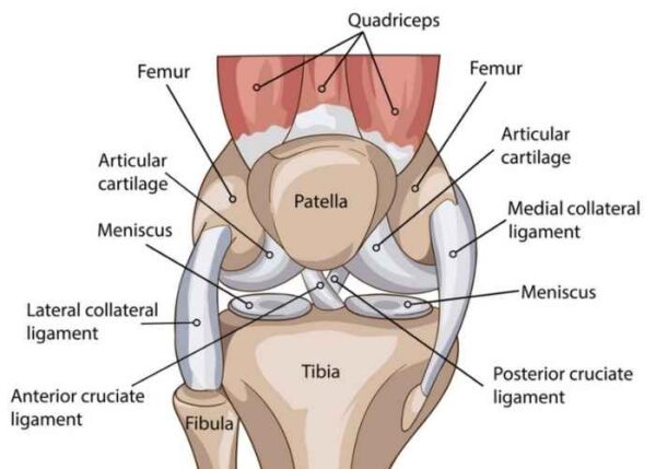
- The thighbone (femur)
- The shinbone (tibia)
- The kneecap (patella)
Another important structure connected to knee anatomy and the three bones is articular cartilage. Articular cartilage is a smooth, hard substance responsible for decreasing knee friction and allowing the bones to glide smoothly against each other as the knee joint bends and straightens. Two wedge-shaped pieces of meniscal cartilage, known as menisci, serve as “shock absorbers” between the femur and tibia. The meniscal cartilage helps to decrease the load on the articular cartilage and prevents development of knee arthritis.
Ligaments act like strong ropes to hold the bones together and provide stability to the knee joint. These ligaments include:
- Anterior cruciate ligament (ACL) – The ACL travels from the anterior of the tibia to the posterior of the femur and prevents the tibia from moving forward in relation to the femur. The ACL is one of the most important structures in the knee and is most commonly injured in movements that require twisting.
- Posterior cruciate ligament (PCL) – The PCL travels from the posterior of the tibia to the anterior of the femur and wraps around the ACL, preventing the tibia from moving backwards on the knee. It is the largest and strongest ligament in the knee joint and is not frequently injured.
- Medial collateral ligament (MCL) – The MCL is a band that runs between the inner surfaces of the tibia and femur and prevents the knee from collapsing inwards.
- Lateral collateral ligament (LCL) – The LCL is located on the outer surface of the femur and fibula on the outside of the knee joint. The LCL resists impact from the knee’s inner surface and prevents the knee from collapsing outwards.
The cruciate ligaments (ACL and PCL) are responsible for controlling the back and forth motion of the knee joint, while the collateral ligaments (MCL and LCL) are responsible for controlling the sideways motion of the knee.
The remaining structures that compose knee anatomy are the tendons and muscles. The quadriceps tendon connects the front thigh muscles to the patella. The patellar tendon stretches from the patella to the shinbone. Both muscles allow the knee joint to extend and become straight.
Since the knee joint is such as complex joint in the human body, it is prone to injuries from a fall, sports collision, overuse, a degenerative condition or automobile accident. Common knee injuries include:
For more information on knee anatomy, or for additional resources on causes of knee joint injuries, please contact the Vail, Aspen, Colorado Springs and Denver, Colorado orthopedic office of knee specialist Dr. Matthew Provencher.